|
NEUROVASCULAR COMPRESSION |
|
|||||||||||||||||
|
|
|
|||||||||||||||||
|
RESEARCH |
|
|||||||||||||||||
|
|
|
|||||||||||||||||
|
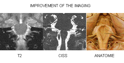
The relationships between the cranial nerves
and the vessels at the surface of the brainstem display a complex three
dimensional (3D) formation. For a number of entities like the neurovascular compression
syndromes or space occupying processes in the posterior fossa and the
reqiured surgical procedures the spatial understanding of these relationships
is very important. The depiction of the relationship between vessels and
cranial nerves predominately was limited to a two dimensional (2D)
presentation. A reproducable and comprehensible assessment was not possible
untill now.
The neurovascular compression is a pathologic contact between a
vessel and a cranial nerve. A series
of entities are caused by such a pathologic contact. The knowledge about the
neurovascular relationships is of great importance. An etiologic connection
of neurovascular compression at the ventrolateral medulla to arterial
hypertension is discussedand and for this the possibility of it’s
demonstration is substantial. The relevant structures are very small and a
proper assignment requires great experience. 3D-visualization requires a
segmentation of the tiny vascular and neural structures. A purely explicite
segmentation of these small structures is extremly time consuming and results
into dissatisfying results of the visualization.
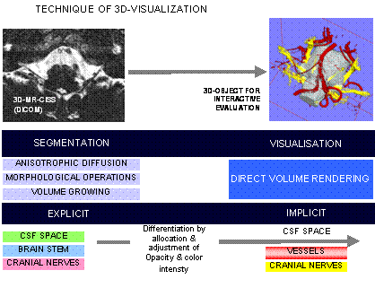
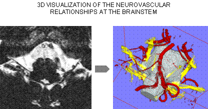
In this project 3D-representations of the
neurovascular relationships at the surface of the brainstem are generated
based on multy modal image data by methods of image and graphic data
processing. Two different strategies are embarked at the same time. On
strategy is based on the improvment and automatization of morphological filtering and volume growing.
This has been already well proven in a semiautomatic approach. The other
strategy is based on nonlinear registration between an anatomic atlas and the
anatomic volume data (
The developed methods are used durind
microvascular decompression of the neurovascular compression syndromes. For
the intraoperative integration the 3D-presentations are provided to the
surgeon within the sterile area. By this the surgeon is able to look at
standard projections as well as to projection coresponding to the
microsurgical view. Furthermore this provides an intraoperative virtual
endoscopy with views of the operative anatomy and regions which are not
visible by the coaxial direction of the operative microscope. The
association of neurovascular compression and arterial hypertension is being
investigated in cooperative scientific study of the department of neurosurgery
and the medical clinic IV - nephrology and the department of gynocolgy of the
University Erlangen-Nuremberg. The neurovascular compression at the
ventrolateral medulla in arterial hypertension is evaluated by the above
presented modern methods of imaging and image processing. In addition the
assossiation between hypertension during pregnancy and neurovascular
compression is evaluated. In a surgical study the possibility and the
effectivness of microvascular decompression for the
treatment of resistant arterial hypertension is under evaluation. This is the
world wide first prospective clinical trial on this subject. It was possible
to normalise or improve the blood pressure in a series of patients. |
COOPERATIONS Frauenklinik
Universität Erlangen-Nürnberg (Dr. Goeke) Neurologische Klinik Universität Erlangen-Nürnberg (Prof. E. Lang) Institut für
Physiologie Universität Erlangen-Nürnberg (Prof. Messlinger) Institut für Graphische
Datenverarbeitung Universität Erlangen-Nürnberg (Prof. Greiner) Neurochirurgische
Klinik Universität Nimwegen, Holland.
(Dr. J. deVries, Dr T. Menovski) |
|||||||||||||||||
|
|
|
|||||||||||||||||
|
NVCHOME.COM |
|
|||||||||||||||||
|
R. NARAGHI |
|
|||||||||||||||||
|
|
|